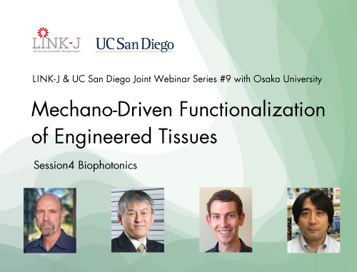
LINK-J & UC San Diego Joint Webinar Series #9 with Osaka University "Mechano-Driven Functionalization of Engineered Tissues" - Session 4: Biophotonics was held on Dec 3, 2021. (Host: LINK-J, Co-Host: University of California San Diego [UC San Diego])
For the ninth in the Joint Webinar Series, LINK-J and UC San Diego were pleased to offer a joint webinar, in cooperation with the Osaka University Global Center for Medical Engineering and Informatics (MEI Center), at which Assistant Professor Johannes Schoeneberg from UC San Diego and Professor Katsumasa Fujita from Osaka University presented recent work in "Biophotonics."
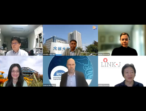
Professors Andrew McCulloch (Distinguished Professor of Bioengineering and Medicine; Director, Institute of Engineering in Medicine, UC San Diego) and Masahiro Kino-oka (Deputy Director, Global Center for Medical Engineering and Informatics; Professor, Department of Biotechnology, Graduate School of Engineering, Osaka University) moderated a discussion.
UC San Diego and Osaka University have a long history of collaborations in education and research. In recent years, they have strived to expand their areas of collaboration to include bioengineering, bioinformatics, tissue engineering, and regenerative medicine.
The recording of the webinar is available from the link below.
https://youtu.be/6fgl2388b9E
Please note that some content is unavailable, and that the webinar might be deleted from the archive in the future.
We encourage you to subscribe to LINK-J's YouTube channel to view great content.


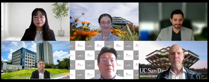
At the beginning of the webinar, Yoshinori Kiuchi (General Manager, LINK-J), Ms. Miwako Waga (Director of International Outreach, UC San Diego), Professor Andrew McCulloch, and Professor Masahiro Kino-oka gave the opening remarks.

"4D Cell Biology: Adaptive optics lattice light-sheet imaging and AI powered big data processing of live stem cell-derived organoids."
Dr. Johannes Schoeneberg (Assistant Professor, UC San Diego School of Medicine)
He introduced his recent studies from the below-mentioned views.
New advanced microscopy methods such as adaptive optics lattice light-sheet microscopy allow fundamentally new insights into cell biology. At the same time, they require high performance computing and machine learning for data processing. The combination allows for four dimensional (4D) cell biology.
To learn more details, please watch the video recording.

"Raman microscopy as a complementary tool for biological imaging"
Dr. Katsumasa Fujita (Professor, Department of Applied Physics, Osaka University)
He introduced his recent studies from the below-mentioned views.
Raman spectroscopy has been an important tool for scientists because of its ability to analyze materials in a label-free manner. Raman spectra reflect the vibrations of molecules in a sample and provide rich information about the composition and environment of the sample.
However, due to the low efficiency of Raman scattering, it has been difficult to use Raman scattering for imaging biological samples under physiological conditions. We have developed a Raman imaging technique that achieves fast Raman imaging by detecting Raman spectra of the sample in parallel. By detecting hundreds of Raman spectra from the illumination line in parallel, we were able to drastically reduce the image acquisition time. We applied this line-illumination Raman imaging technique to label-free observation of molecular dynamics in cellular phenomena such as apoptosis, drug response, cell division, and cell differentiation. 532nm laser excitation is used to monitor mitochondrial dysfunction at the cellular level using the resonance Raman effect of heme proteins. We have also proposed and demonstrated the use of alkynes as tiny tags for imaging small molecules. This enabled us to observe molecules that are too small to be labeled by fluorescent probes.
To learn more details, please watch the video recording.

This webinar included time for Q&A and panel discussion, which led to interesting interactions among the panelists.
Thank you very much for your active participation in the joint webinars. We plan to offer similar sessions in the future. Stay tuned!