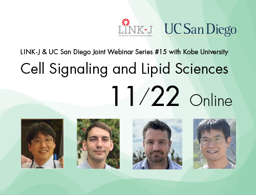
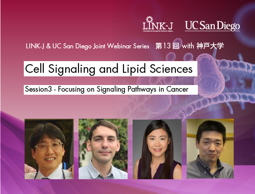
LINK-J & UC San Diego Joint Webinar Series #10 with Kobe University "Cell Signaling and Lipid Sciences" Session 1 - Focusing on Lipid Membranes was held on September 7, 2022. (Host: LINK-J, Co-Host: University of California San Diego [UC San Diego])

Speakers
Dr. Neal K. Devaraj, Professor of Chemistry and Biochemistry, UC San Diego
Dr. Toshiki Itoh, Professor, Biosignal Research Center, Kobe University
Dr. Yasuhito Shirai, Professor, Graduate School of Agricultural Science, Kobe University
Dr. Itay Budin, Assistant Professor of Chemistry and Biochemistry, UC San Diego
Ms. Miwako Waga, Senior Director of International Outreach, UC San Diego
Akihiko Soyama, Chief Executive Officer, LINK-J
Presentations
"Bioconjugation strategies for revealing the roles of lipids in living cells"
Dr. Neal K. Devaraj, Professor of Chemistry and Biochemistry, UC San Diego
Our laboratory developed and applied different bioconjugation strategies to study the role of lipids and lipid modifications in cells. Inspired by our ongoing work on developing lipid bioconjugation strategies to generate artificial cell membranes, we developed a ceramide synthesis method in live cells using a salicylaldehyde ester that readily reacts with sphingosine in form of a traceless ceramide ligation. Our study not only confirmed existing knowledge about the association of ceramides with cell death, but also gave interesting new findings about the structure-function relationship of ceramides in apoptosis. Our initial efforts led us to investigate probes that detect endogenous sphingolipids using live cell imaging. We describe the development of a fluorogenic probe that reacts chemoselectively with sphingosine in living cells, enabling the detection of elevated endogenous levels of this biomarker in human disease. Building on our interest in the fluorescence labeling of lipids, we have also explored the use of bioorthogonal reactions to label chemically synthesized lipid probes. I will discuss the development of photocaged dihydrotetrazine lipids, where the initiation of the bioorthogonal reaction can be triggered by visible light, allowing for live cell modification of membranes with spatiotemporal control.
"Membrane tension and membrane-bending proteins in the regulation of cell migration"
Dr. Toshiki Itoh, Professor, Biosignal Research Center, Kobe University
Cells are not simply spherical in shape, but have a variety of shapes according to their functions. When we draw a picture of a cell on paper, the outline usually corresponds to the cell membrane. In other words, the basis of the diversity of cell morphology is the mechanism that controls the shape of the cell membrane. The plasma membrane is subjected to a physical force called membrane tension, which is the sum of (1) the planar tension that pulls the lipid bilayer horizontally and (2) the adhesive force between the plasma membrane and cortical actin. The former is thought to be contributed mainly by the osmotic pressure difference between the inside and outside of the cell, while the latter is thought to be contributed mainly by the interaction between plasma membrane phospholipids and actin-binding proteins. We have identified the F-BAR domain, a conserved region with membrane-deforming activity, and are analyzing its physiological function. The F-BAR domain is conserved in FBP17, which activates the actin polymerization factor N-WASP-Arp2/3, Slit-Robo GAPs (srGAPs), which are RhoGAPs, and Fer and Fes, which are tyrosine kinases. Therefore, F-BAR domain-containing proteins are thought to functionally link actin polymerization to the shape of the plasma membrane. However, the specific mechanism of how the membrane deformation activity of the F-BAR domain regulates actin polymerization in cancer cell invasion and other processes has remained unclear. We recently found that the membrane deformation activity of the F-BAR domain functions as a sensor of plasma membrane "bendability," or membrane tension. Actin polymerization increases plasma membrane tension, and increased membrane tension causes F-BAR domain proteins to dissociate from the plasma membrane. Thus, the parameter of plasma membrane shape provides a scaffold for actin polymerization and may also regulate cell motility through a feedback mechanism mediated by membrane tension.
This webinar included time for Q&A and panel discussion, which led to interesting interactions with the audience. Thank you very much for your participation. We plan to offer similar sessions in the future. Stay tuned!